Norwegian Rune Brautaset discovered his interest in research when writing his BSc thesis in England in 1998 during his optometric training.
There was something I had never experienced before: a prominent, interdisciplinary and active research environment in optometry, in basic research as well as clinical research and implementation, which really appealed to me, says Rune Brautaset, researcher and Senior Lecturer, Head of the Division of Eye and Vision in the Department of Clinical Neuroscience at Karolinska Institutet located at St. Erik Eye Hospital.
In 2000 he started working as a lecturer at Karolinska Institutet (KI) and three years later, he defended his thesis. The love of his Swedish wife made Rune Brautaset to remain in Sweden and with inspiration from England, he helped build the optometric research and education currently available at KI and St. Erik Eye Hospital.
I am driven by curiosity and by helping people have better lives. At the same time, I find research very stimulating and somewhat competitive, which suits me.
Since 2018, he is the Head of Division of Eye and Vision at KI’s Department of Clinical Neuroscience. The division, which is located at St. Erik Eye Hospital, provides education and conducts research within ophthalmology and optometry as well as eye nurse training. The teaching clinic receives around 3,500 patients per year.
Researches OCT analysis
Today, Rune Brautaset main research interest lies in the structure and function of the eye, in which the technology called optical coherence tomography (OCT) is essential. Even if the OCT instrument itself costs around SEK 500,000, OCT can be used to easily, quickly and inexpensively examine the different layers of the eye’s structure, such as the retina nerve layers.
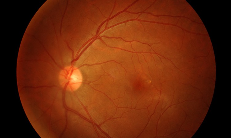
The retina in the eye photographed with OCT.
Photo: Istock
Rune Brautaset’s research team includes programmers that can conduct more advanced analyses of the OCT images than what the instrument can do on its own. The research has primarily involved OCT and the disease MS, which is a chronic, neurological and autoimmune disease affecting the brain and the spinal cord.
The retina is a window to the brain
MS patients usually undergo costly and time-consuming magnetic resonance imaging (MRI) examinations to measure the nervous tissue of the brain and spinal cord.
In the case of MS, the retina of the eye can be seen as a window to the brain.
Using OCT, we can discover and monitor the progress of disease. In MS, the loss of nervous tissue that the patient has in the brain is reflected in the retina of the eye and we can see if a certain treatment is slowing the progress of the illness. It is also easier to measure the changes in the nervous tissue of the eye in MS, as it does not become inflamed and swollen in the same way as it does in the brain, says Rune Brautaset.
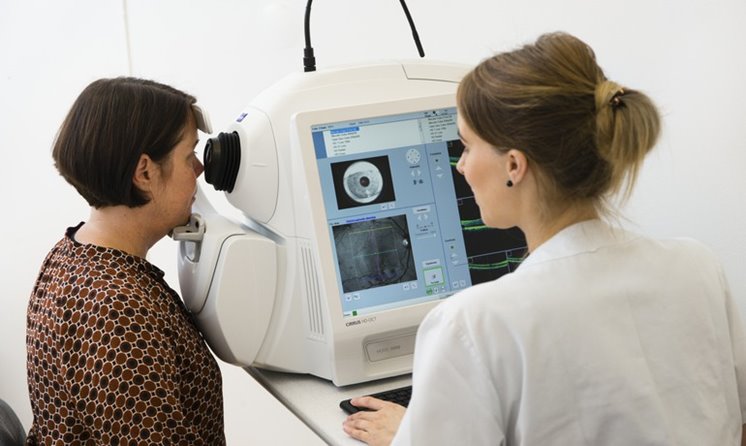
A patient during an OCT-examination.
Photo: Johanna Hanno
When the research team gathered a large group of MS patients in a study, they drew the conclusion that if you use the same brand of OCT instrument for each examination of a patient, it can replace some of the MRI examinations. The reason is that using the same brand of OCT machine allows for a correct long-term interpretation of the data analysis.
The research team is now continuing to refine the analyses and to collaborate with other teams to use OCT in the examination and monitoring of more diseases, including HIV and Alzheimer’s disease.
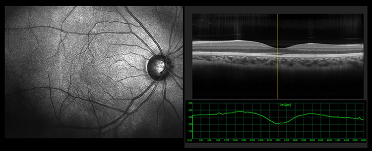
OCT images of the retina and its nerve layers.
Photo: Istock
Problems with binocularity can be mistaken for dyslexia and ADHD
In the first ten years of his career, Rune Brautaset focused on binocular vision; “why we see one image with two eyes”, and problems associated with binocularity. When your binocular vision works as it should, the eye movements are coordinated, so that the images seen by both eyes can be merged into one, and you can focus and achieve a high level of visual acuity.
If the binocularity is not working, the affected person may unconsciously reread the same line, feel that the text is moving, quickly tire of reading, read slowly or have trouble understanding what a text is about. You can also develop headaches as well as blurred or double vision. The cause can be a refractive error or that the interior or exterior muscles of the eye is not functioning properly.
It is important to receive the right diagnosis based on the symptoms and coordination measurements. Otherwise, there is a risk of mistaking binocularity problems with dyslexia (reading and writing difficulties) or ADHD (a neuropsychiatric functional impairment) as the behaviour in a reading situation will be partially identical.
Research indicates that five to ten per cent of children have these issues and that the proportion needing help is increasing. They do not need medical care, but help with eyesight training methods and corrective glasses, says Rune Brautaset.
Problems grow along with increased near work
Binocularity issues usually manifest once children go from learning how to read to reading for the purpose of learning, i.e. in third or fourth grade. They also appear when the amount of near work increases later in life.
International studies also show that binocularity issues are more common among school children with ADHD than in other children. It aggravates the child’s ADHD, as reading issues leads to exclusion. However, binocularity issues can often be helped with exercises, and if more children had the opportunity to do those exercises we would have a more quiet school environment where more young people are happy and increase their level of knowledge.

Binocularity issues usually manifest when the amount of near work increases.
Photo: Istock
In his research, Rune Brautaset has evaluated the type of traditional binocular training that non-squinting children with binocularity issues have been given for nearly 150 years.
The most important conclusion is that doing exercises for 16 weeks has a short-term effect, which dissipates when you stop doing them. But if you continue the exercises around 16 more weeks after the symptoms have disappeared, you will have a long-term effect. That’s where you need to find the motivation to keep going, he says.
Patented technology for binocularity training
In 2008, Rune Brautaset patented a new, unique treatment method that he developed for binocularity training using a computer, which is intended to keep people from giving up their exercises when the symptoms disappear.
Then you can watch a movie or play a computer game at the same time as doing your visual exercises without even noticing, due to the manipulation of the image. You get a larger volume of exercise, while the treatment provider can monitor and alter your exercises, he says.
The treatment, which entails the patient renting a computer and screen from their optician, is costly since it is not subsidised by the government.
Countless patients could receive help if we are able to develop and offer the training programme at a lower cost through an app. Then you could plug your mobile into a pair of VR glasses and do your exercises. We are therefore looking for funding and partners to adapt the solution to mobile uses.
Text: Helena Mayer